Introduction
The temperomandibular joints area is challenging for good diagnostic
radiographs. It is a common area of injury, particularly in cats.
Failure to diagnose luxations or fractures quickly, following trauma,
can make treatment very difficult later.
|
TMJ Dorso-ventral
Place the patient in sternal recumbency and place pads around the
head, to keep the hard palate parallel to the table.
Place the film on the table for a true DV skull view.
There is much superimposition with this view, but both the mandibular
condyles can be viewed and their position assessed as normal or
not.

|
|
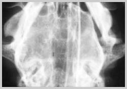
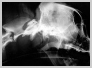
DV view of caudal dog skull clearly showing
right mandibular condyle luxated rostrally out of the TMJ. Compare
with normal left side
|
| |
|
|
|

DV view of caudal dog skull clearly
showing both mandibular condyles within the TMJ's
|
|
|
|
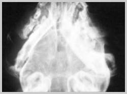
DV view of caudal cat skull showing
neoarthrosis of both mandibular condyles
|
|
TMJ Sagittal Oblique
|
|
This is a useful view for the temperomandibular joint spaces.
The patient is placed in lateral recumbency with the joint to be
examined nearest the table. The rostral aspect of the head is raised
so that the sagittal plane is raised rostro-caudally by 25°
for brachycephalic breeds, 15° for mesocephalics and 10°
for dolicocephalics.The mouth is opened with a foam block and the
central beam is angled onto the joint to be examined.
Try to align the long axis of the mandibular condyle perpendicular
to the film for better visualisation of the joint space.
|
|
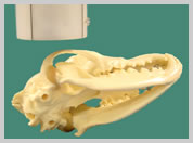
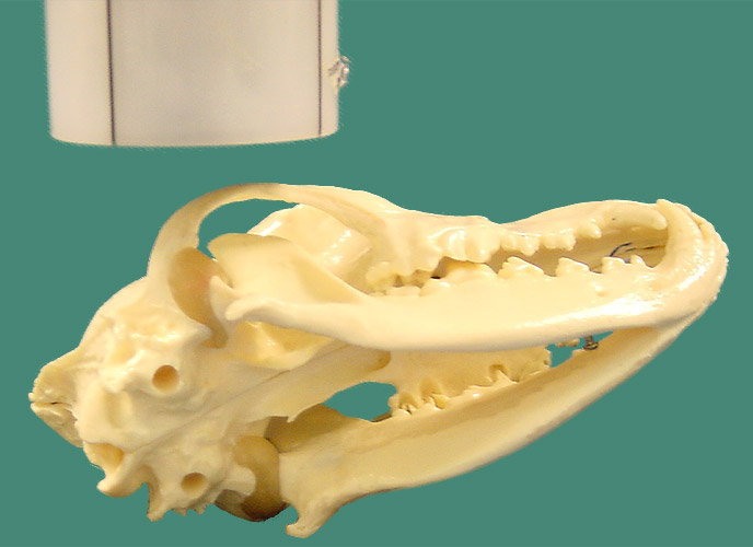
Position for TMJ sagittal oblique
view with plate on table and joint to be imaged closest to
the plate. Note direction of beam down long axis of condyle
|
|
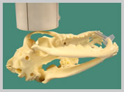
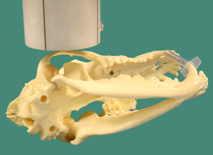
Position for TMJ sagittal oblique
view with plate on table and joint to be imaged closest to
the plate - mouth open
|
|
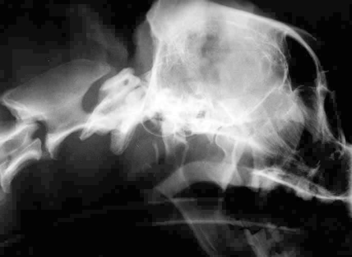
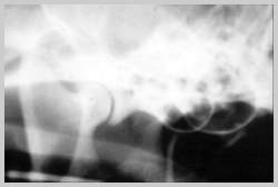
Radiograph showing joint space using open mouth technique
variation |
|
| |
| |
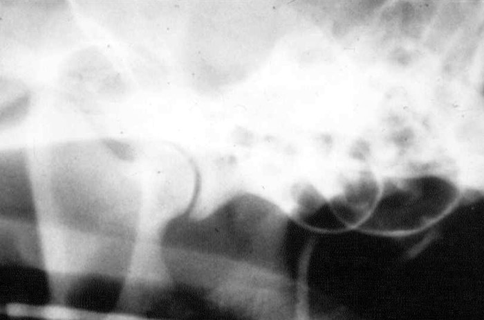
Radiograph showing joint space using above technique
|
|
|
| |
TMJ Lateral Oblique Views - Cat
Due to the prominence of the zygomatic arch in the cat, a slightly
different view is employed - the ventro 20° lateral-dorsolateral
oblique.
The patient is placed in lateral recumbency, with the target joint
away from the table. The head is tipped up 20° from the lateral
plane and the beam is directed perpendicularly through the upper
TMJ.
| |
Position for cat TMJ's avoiding the zygomatic arch. Note that
the joint to be imaged is away from the film
|
|
|