Introduction
Recently, the greater availability of CT scanners has allowed this
imaging method to be used, to complement the usual radiographic
techniques, for some of the more complex conditions.
|
|
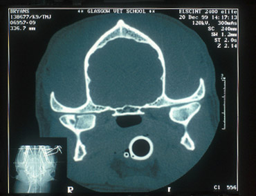
CT scan of fractured right mandibular
condyle
X-ray & CT images
courtesy of Glasgow University
|
|
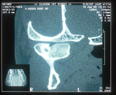
CT scan of same fractured mandibular condyle 4 weeks post-trauma
|
|
|
|

Cat having a CT scan at Glasgow
Veterinary School
|
|
Advantages
- Provides powerful images and computer reconstruction of
otherwise hard to examine areas, such as TMJ's, caudal mandibular
body, coronoid process and zygomatic arch
- Provides a 3D image and reconstruction of neoplasms with
likely surgical margins within the cranium, nasal cavity
and orbital area
- In time, it may prove more user-friendly than radiographs
for imaging the roots of horse and other herbivore teeth.
Disadvantages
- Cost
- Complex machines to use without specialist training
- The image quality is substantially poorer than non-screen
and dental radiographs for detail.
|
|
|
|
|