|

|
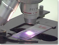
Slide Examination
Try to adopt a methodical approach. The following routine is satisfactory
in most cases.
- Examine the specimen on the slide by eye. Evaluate slide preparation
e.g. is the staining adequate. Occasionally you will see a recognisable
object e.g. louse egg, large mite, nymphal tick
- Start with the lowest low power objective. If you can easily
see the hair shafts, you will be able to recognise mites and other
parasites. Do not use a higher power for parasites
|
|
 |
|
|
The paradox: The time required for examination
increases by the square of the increase in magnification.
Changing from a x 2 to a x 10 objective implies a 25-fold
increase in the time required to make the same search!
You do not have the time so you make a less thorough
search.
|
|
|
| |
|
- Scan the slide methodically with a pattern, which covers the
whole of the specimen. Use higher power for anything, which is
not clear at low power. Mover the coverslip or remount to expose
objects of interest which are obscured
- For cytology e.g. tape strips, scanning quickly at low power
is still useful. You may spot something unexpected e.g. Demodex
in pus, and you can identify suitable areas of the preparation
for high power examination i.e. well stained, not too thick or
thin
|
|
|
|
|
