Culturing for Malassezia
Whilst cytology is the main method for demonstrating and enumerating
Malassezia in skin lesions, cultures can also be of value.
Routine swabbing methods are appropriate in the ear and from greasy
skin lesions.
Sabouraud's dextrose agar supports the growth of M. pachydermatis
from dogs; plates should be incubated at 32-37°C for 3-7 days.
M. pachydermatis produces domed or high convex, entire, yellow-buff
colonies. Lipid supplemented media such as modified Dixon's agar
are required for the isolation of other Malassezia spp. which
are of lesser importance in other veterinary species.
Click to zoom
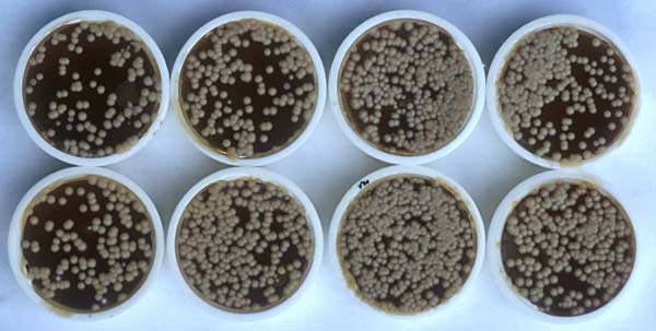
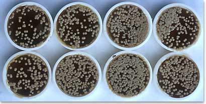
Contact plates containing modified Dixon's
agar
Note the numerous yellow M. pachydermatis colonies indicating high
yeast population densities on the skin
Contact plates are a useful alternative and provide a useful guide
to population densities of the yeast (see above). Small bottle lids
are filled to the brim with medium and then simply applied to the
skin surface for 10 seconds, removed, then incubated. Colonies can
be counted after incubation to give an impression of the population
density.
|