
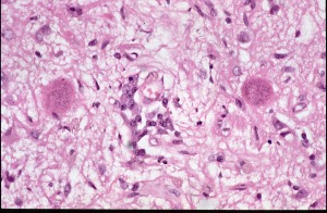
Section from brain of a dog infected with Toxoplasma gondii
This is a low power view of a section from the brain of a dog infected with T.gondii. The dog had a history of fits and convulsions and on necropsy large numbers of T.gondii cysts were found in its brain.
The two pink bodies with darker staining granules are the tissue cyst forms of the parasite and contain large numbers of bradyzoites - the slow growing stage of the parasite. The bradyzoites are the infective stage for the final host - a carnivore and would infect anything that eat the dog.
The dog itself probably became infected through eating meat containing similar tissue cysts. Notice the absence of any cellular reaction around these cysts.
See also