
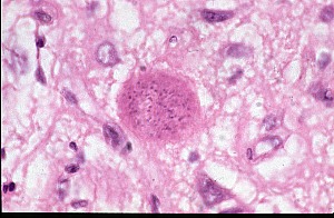
Section from brain of a dog infected with Toxoplasma gondii
This is a high power view of a section from the brain of a dog infected with T.gondii. The dog had a history of fits and convulsions and on necropsy large numbers of T.gondii cysts were found in its brain.
The large rounded pink body is a the tissue cyst form of the parasite and has a very thin outer wall. The darker staining granules inside the cyst are the nuclei of and the individual bradyzoites - the slow growing stage of the parasite.
The bradyzoites are the infective stage for the final host but sometimes the bradyzoites become activated in the host e.g. due to changes in the immune system which leads to disease localised within the affected host organ. The brain is a common site for tissue cysts and reactivation of bradyzoites is often associated with CNS dysfunction.
See also